

Imagine a regular morning that suddenly feels off. One eye aches when you move it, colors look strangely muted, and your central vision seems like you’re looking through smudged glass. It’s unsettling, and for many people, this is exactly how optic neuritis first announces itself. Not a headache, but not blurry vision from screen fatigue either. Something different.
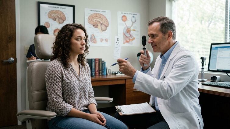
This is not a standard vision problem that a new glasses prescription will fix. It is a neurological condition, and it needs the right specialist. At Lone Star Neurology, we have been caring for patients across Texas for over 19 years. We know that early recognition changes everything with this condition. Here is what you need to understand.
What Optic Neuritis Is and How It Affects Vision
Your optic nerve contains over a million fibers responsible for transmitting visual signals from the retina to the brain. When inflammation strikes, those signals get distorted, delayed, or blocked entirely.
Optic neuritis vision loss works through a fundamentally different mechanism than conditions like glaucoma, where the culprit is pressure inside the eye. Here, the damage is inflammatory. The myelin sheath (the protective insulating layer wrapped around nerve fibers) begins to break down. When that insulation deteriorates, nerve impulses slow down or misfire. The result: blurred images, reduced contrast sensitivity, and colors that look washed out. Red is almost always the first to be affected, shifting toward brown or gray in the involved eye.
Understanding this mechanism matters clinically. You are not dealing with a lens problem or a retinal tear. You are dealing with the nervous system, which is exactly why a neurologist, not just an ophthalmologist, needs to be part of your care team from the start.
Symptoms of Optic Neuritis
Optic neuritis symptoms tend to develop gradually, over hours or days, which leads many people to wait, hoping things will improve on their own. They often don’t, and waiting costs time that matters.
Pain with eye movement is the most hallmark feature. It can range from a dull ache to a sharp stabbing sensation, and it consistently worsens when you look to the side or up and down. One of our patients described it as “feeling like my eye was being pulled from the inside every time I moved it.” That description is more accurate than it sounds.
Vision becomes foggy or dimmed – many patients say it feels like looking through frosted glass. Optic neuritis vision at this stage often fluctuates: symptoms tend to worsen with heat or physical exertion and partially ease with rest, a phenomenon called Uhthoff’s sign that neurologists specifically look for.
Optic neuritis headache is a common accompanying complaint, typically localized around or behind the affected eye. Patients with a pre-existing migraine history sometimes initially attribute this to a migraine episode – a distinction our headache specialists at Lone Star Neurology are specifically trained to evaluate.
Color desaturation, particularly involving red tones, rounds out the classic presentation of optic neuritis symptoms. If a red stop sign looks faded or brownish through one eye, that is a real clinical sign, not imagination.
What Causes Optic Nerve Inflammation
The most common driver is autoimmunity: the immune system mistakenly targets the myelin sheath of the optic nerve and begins breaking it down. Optic neuritis causes include multiple sclerosis, neuromyelitis optica spectrum disorder (NMOSD), and systemic autoimmune conditions like lupus or sarcoidosis. Viral infections can also trigger the inflammatory cascade, sometimes appearing two to four weeks after a systemic illness.
One specific form deserves special attention: retrobulbar optic neuritis – inflammation behind the eyeball rather than at the optic disc. In these cases, the fundus examination can look completely normal, even while the patient experiences significant vision loss and pain. This is exactly where retrobulbar neuritis becomes diagnostically tricky: because the eye looks “fine” on a standard exam, some patients spend weeks being reassured when they should be getting an MRI.
Optic nerve inflammation is also, in many cases, the very first clinical signal that something larger is happening in the nervous system. Which is why the next step is proper diagnosis, and it matters so much.
How Neurologists Diagnose Optic Neuritis
Optic neuritis diagnosis requires a layered approach. A standard vision chart tells you how much the patient has lost. It does not tell you why or what comes next.
At Lone Star Neurology, our neurologists use a comprehensive workup that includes:
- MRI with contrast – the cornerstone of evaluation. It visualizes active inflammation along the optic nerve and, critically, detects white matter lesions in the brain that may indicate multiple sclerosis. If you want to understand what contrast MRI actually shows and why it matters, our blog post on MRI brain imaging with and without contrast clearly walks through the key differences.
- Visual evoked potentials (VEP) – this test measures the speed at which signals travel from the eye to the brain. Slowed conduction confirms myelin damage even when other findings appear borderline.
- Optical coherence tomography (OCT) – a non-invasive scan measuring the thickness of nerve fiber layers in the retina. It provides both a diagnostic snapshot and a baseline for tracking changes over time.
- Blood work – testing for anti-AQP4 antibodies (a marker for NMOSD), MOG antibodies, and general autoimmune markers. Results directly influence long-term treatment decisions.
- Pupillary response (RAPD) – when one optic nerve is damaged, the affected pupil reacts differently to light. A trained neurologist will detect this immediately on examination. To learn more about how the brain and nervous system process these signals, our article on brain anatomy and function provides a solid foundation.
Treatment Options for Optic Neuritis
Standard optic neuritis treatment begins with high-dose intravenous corticosteroids, typically methylprednisolone, administered over 3 to 5 days. The goal is to rapidly suppress inflammation, shorten the recovery period, and reduce cumulative nerve damage. Steroids do not always change the final level of recovered vision, but they meaningfully accelerate the timeline.
Following IV therapy, a course of oral steroids is often prescribed to taper the treatment and lower the risk of early relapse.
In severe cases or when the underlying condition is NMOSD, where attacks tend to be more aggressive and recovery less complete, plasmapheresis is used to remove the abnormal antibodies driving the attack.
In mild cases, symptoms sometimes improve partially on their own. But “may partially resolve” is not a reason to skip monitoring; even low-severity episodes require neurological follow-up to catch any progression of an underlying condition.
Optic Neuritis and Multiple Sclerosis
This is the connection that understandably worries patients most, and it deserves a direct, honest answer.
Yes, optic neuritis is one of the most frequent first presentations of multiple sclerosis. A substantial proportion of patients who experience a first isolated episode will eventually develop MS, particularly when their MRI already shows additional brain lesions at the time of the episode.
The important point: this is precisely why early neurological evaluation changes outcomes. When MS is identified before multiple relapses have occurred, disease-modifying therapy can significantly slow its progression. Our multiple sclerosis center specializes in exactly this kind of early, proactive management, from diagnosis through long-term care.
Not every case of optic neuritis leads to MS. But every case deserves a thorough neurological workup to answer that question with certainty, not guesswork.
Recovery and Long-Term Outlook
Most patients begin noticing visual improvement within 2 to 4 weeks, with recovery continuing for up to 6 months. The majority regain vision close to their baseline – though some residual effects, like reduced contrast sensitivity or subtle shifts in color perception, can persist even after the inflammation resolves.
Long-term management matters just as much as acute-phase management. Recurrence is a genuine risk, especially in patients with autoimmune conditions. Regular neurological follow-up enables our team to detect new episodes early, adjust treatment if the underlying diagnosis evolves, and monitor nerve fiber status with serial OCT imaging.
Patients who stay connected with their neurologist consistently do better, not because their disease is necessarily more severe, but because problems get addressed before they compound.
When to See a Neurologist About Vision Changes
Here is a practical rule worth remembering: if your vision changes suddenly and your eye hurts when you move it, do not wait for an appointment slot to open next week. Call the same day.
Pain with eye movement, combined with sudden visual blurring, signals potential optic nerve inflammation and warrants urgent evaluation. The same applies to sudden loss of color brightness or any rapid dimming in one eye, even if it feels mild or intermittent at first.
At Lone Star Neurology, we offer same-day appointments and operate 18 locations across Texas. Our neurologists handle the full diagnostic complexity these cases require: the imaging, the bloodwork, and the long-term planning that follows. Book an appointment today or call us at 214-619-1910.
FAQ
Can optic neuritis cause permanent blindness?
In most cases, vision partially or nearly fully recovers with appropriate treatment. However, severe nerve damage or significantly delayed care can result in lasting deficits, which is why prompt evaluation is so important.
Is optic neuritis always a sign of multiple sclerosis?
No. While MS is a common underlying cause, the condition can also result from other autoimmune diseases, infections, or inflammatory processes. A thorough neurological workup determines the actual cause.
How long does recovery take?
Initial improvement typically begins within a few weeks. Full recovery may take several months, depending on the severity of nerve involvement.
Can it come back after treatment?
Yes, relapses are possible – particularly in patients with autoimmune or inflammatory conditions. This is why ongoing follow-up with a neurologist remains part of long-term care.
Should I see a neurologist or an ophthalmologist for optic neuritis?
 (No Ratings Yet)


I've given up... the stress her office staff has put me through is just not worth it. You can do so much better, please clean house, either change out your office staff, or find a way for them to be more efficient please. You have to do something. This is not how you want to run your practice. It leaves a very bad impression on your business.
Please, leave your review
Write a comment: