

In neurosurgery, treating cerebral aneurysmata has seen remarkable progress. It is thanks to modern medical advances. This illness is a life-threatening condition characterized by the ballooning of a blood vessel in the brain. It demands swift and sophisticated interventions to prevent rupture and subsequent complications. Advances in medical technology, surgical techniques, and interventional procedures have been significant. They have reformed the area of brain aneurysm treatment. Our guide examines current approaches and innovations. They are shaping cutting-edge treatment. The arsenal of treatment options has expanded. It happens from invasive endovascular procedures to cutting-edge neurosurgical interventions.
They offer patients more choices and improved outcomes. Understanding these modern advances is crucial for healthcare professionals. It empowers individuals and their families with severe knowledge. They can guide informed decisions in navigating the complexities of brain aneurysm treatment. Join us on a trip through the latest breakthroughs. Here, science and technology converge to pave the way for more effective and patient-centric approaches.
Diagnosis of Brain Aneurysm
In neurology, the early and accurate diagnosis of cerebral aneurysmata is paramount. It helps for effective intervention and prevention of potentially devastating consequences. Detecting an aneurysm in the brain involves a comprehensive process. It is often utilizing advanced imaging technologies and clinical assessments. This exploration navigates the intricacies of brain aneurysm diagnosis. We shed light on the evolving methods employed by healthcare professionals. Understanding these diagnostic approaches is crucial. They lay the foundation for timely and precise medical interventions. They also provide insight into advances in neuroscience in the quest to improve patient outcomes.
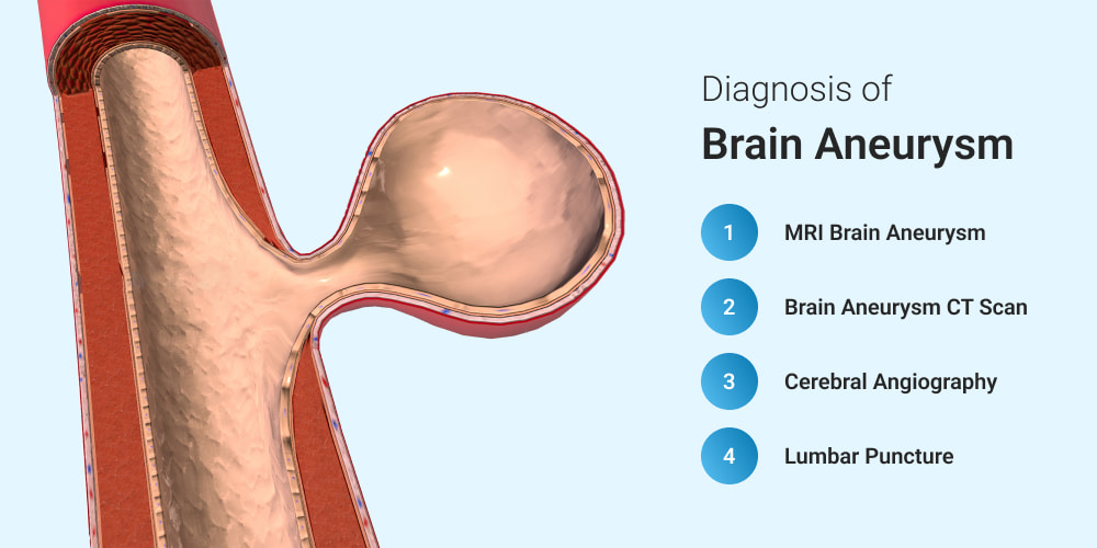
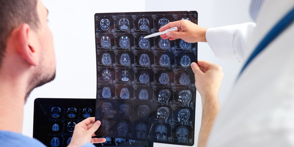
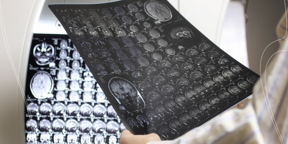
MRI Brain Aneurysm
This technique offers detailed insights into cerebral anatomy in MRI brain aneurysm diagnosis. Here’s a list outlining the aspects of using it:
- High-Resolution Imaging: MRI provides high-resolution images, aiding in detecting small aneurysms.
- Multi-Planar Views: It allows for multi-planar views. Also, it facilitates a comprehensive assessment of aneurysm morphology.
- Tissue Characterization: MRI helps characterize the surrounding tissues. It offers valuable information about potential complications.
- Contrast-Enhanced MRI: Contrast agents can enhance visualization, clearly highlighting aneurysm features.
- Non-Invasiveness: MRI eliminates the need for invasive procedures, providing a safer diagnostic option.
- Evaluation of Blood Flow: These sequences assist in evaluating blood flow dynamics with aneurysms.
- Patient Safety: MRI is safe for most patients. It avoids exposure to ionizing radiation associated with other imaging modalities.
Using these advantages, MRI brain aneurysm diagnosis is a powerful and versatile tool for physicians. It helps in the evaluation and treatment of this complex vascular condition.
Brain Aneurysm CT Scan
A CT scan is a pivotal diagnostic tool offering valuable insights into the intricate vascular area of the brain. Here’s a comprehensive exploration of the critical aspects. They surround the use of CT scans in diagnosing brain aneurysms:
- Rapid Imaging: It provides swift imaging of the brain’s vascular structures. Also, CT scans enable quick assessment.
- Non-Invasive Nature: The procedure is non-invasive. It avoids the need for catheters or contrast agents.
- Detection of Bleeding: Brain aneurysm CT scan effectively detects signs of bleeding. They are crucial in identifying a ruptured aneurysm.
- Enhanced Imaging Technology: Advancements in CT technology, such as CTA, offer detailed 3D images. They enhance diagnostic precision.
- Risk Stratification: These scans aid in assessing the risk of an aneurysm rupture based on size, location, and shape.
- Emergency Situations: CT scans are instrumental. They help to evaluate and manage potential aneurysm ruptures in emergencies.
- Follow-up monitoring: It plays a role in post-treatment monitoring. It ensures the aneurysm’s stability after treatment.
Utilizing the capabilities of a CT scan is pivotal. So, healthcare professionals can swiftly and accurately diagnose aneurysm. It enables timely intervention and personalized treatment strategies.
Cerebral Angiography
This method stands as a vital diagnostic tool in the intricate process of diagnosing brain aneurysms. This invasive yet exact procedure involves injecting contrast. It is into blood vessels to visualize the arteries in the brain. Here is a comprehensive list detailing the aspects of cerebral angiography in diagnosing aneurysms:
- Contrast Dye Injection: It highlights blood vessels. Also, such injection produces detailed images of the cerebral arteries.
- Visualization of Blood Flow: Cerebral angiography enables real-time visualization of blood flow. It helps identify abnormalities, including aneurysms.
- Location Identification: It precisely pinpoints the location of an aneurysm. Local identification aids in treatment planning.
- Size and Shape Assessment: The procedure offers detailed information on the size and shape of the aneurysm. It is crucial to determine the potential risk of rupture.
- Guidance for Treatment Decisions: The detailed images obtained assist doctors in making decisions. They are about the most suitable treatment approach.
- Evaluation of Surrounding Structures: It allows us to test the effect of the aneurysm on nearby structures. Also, cerebral angiography contributes to a comprehensive diagnosis.
- Dynamic Imaging: The dynamic nature of the procedure captures the blood flow in real-time. It offers a more thorough understanding of the vascular anatomy.
- Precise Catheter Navigation: Precise catheter navigation provides targeted visualization of specific blood vessels. It enhances diagnostic accuracy.
Cerebral angiography can provide detailed and dynamic images. It plays a crucial role in the precise and accurate diagnosis of aneurysms.
Lumbar Puncture
Explore the multifaceted role of such punctures in diagnosing brain aneurysms. It underscores its significance in assessing and understanding this complex medical condition:
- Procedure Overview: This puncture involves inserting a needle into the spinal canal to extract CSF for analysis.
- Indirect Imaging Aid: Lumbar puncture is not a direct imaging technique. It indirectly helps diagnose cerebral aneurysms by assessing the CSF for signs of bleeding.
- Detection of Rupture: Elevated red or white blood cell counts and xanthochromia in the CSF may indicate a ruptured aneurysm.
- Complementary to Imaging Studies: Lumbar puncture complements advanced CT and cerebral angiography. They offer additional insights into aneurysm presence and severity.
- Invasive yet Informative: Lumbar puncture provides valuable diagnostic info despite being invasive. It contributes to a more comprehensive understanding of brain aneurysms.
Recognizing the role of lumbar puncture in diagnosing aneurysms is crucial. So, healthcare professionals can integrate this technique judiciously into the diagnostic process. It enhances the overall accuracy and effectiveness of the assessment.
Brain Aneurysm Treatment
The area of brain aneurysm treatment has evolved significantly. It incorporates a range of approaches to address this complex medical condition. The choice of treatment depends on factors such as the size and location of the aneurysm, the patient’s health, and the risk of rupture. Selecting the most appropriate aneurysm healing involves a collaborative decision-making process. It is between healthcare professionals and patients. Such a process considers the specific characteristics of the aneurysm and the individual’s health. Advances in technology and medical techniques continue to shape the sector. It offers a range of options to enhance patient outcomes.
Brain Aneurysm Surgery
This surgery stands as a significant intervention. It is mainly for aneurysms at risk of rupture. Advances in neurosurgical techniques have transformed the area of brain aneurysm surgery. It offers improved outcomes and reduced risks.
- Clipping: A traditional approach is clipping. It involves placing a metal clip around the neck of the aneurysm to prevent rupture. This method remains effective and is often utilized for specific aneurysm types.
- Coiling (Endovascular Embolization): A less invasive option is coiling. It involves inserting a catheter through the blood vessels into the aneurysm using removable coils. The coils induce clotting, sealing off the aneurysm and reducing the risk of rupture.
- Flow Diverters: A more recent innovation is flow diverters, which are stent-like devices. They are often placed across the aneurysm’s neck and redirect blood flow. It promotes the natural healing of blood vessels and prevents their rupture.
- Pipeline Embolization Device: A specific type of flow diverter, PED, diverts blood flow away from the aneurysm. It encourages blood vessel reconstruction.
The decision between these techniques depends on the aneurysm’s size and location. Also, it relies on the patient’s overall health. Advances in brain aneurysm surgery techniques have significantly improved outcomes. Many procedures now offer reduced recovery times and enhanced patient safety.
Brain Aneurysm Treatment Without Surgery
Surgical intervention is a common approach for addressing brain aneurysms. There are alternative methods for healing without surgery. These noninvasive options are often considered for aneurysms that pose a lower risk of rupture. They are also observed in cases where surgery may lead to higher potential complications. Critical brain aneurysm treatment without surgery includes:
- Observation: Monitoring the aneurysm through regular imaging studies. It may be a viable approach for some patients, especially if it is small and stable.
- Medication: Blood pressure-lowering drugs can help manage the risk factors. Also, they may reduce the likelihood of rupture.
- Endovascular Procedures: Catheter placement or stent placement still involves catheter-based procedures. They aim to treat the aneurysm from within the blood vessels.
- Lifestyle Modifications: Healthy lifestyles include managing blood pressure and avoiding smoking. Also, it involves maintaining a balanced diet and regular exercise. It can contribute to overall vascular health and reduce the risk of aneurysm progression.
- Embolization: This procedure involves injecting materials into the aneurysm to induce clotting. Also, it helps to prevent blood flow, reducing the risk of rupture.
The choice between surgical and non-surgical approaches depends on various factors. They include aneurysm size, location, and the patient’s overall health. Exploring brain aneurysm treatment without surgery is vital. So, healthcare professionals can tailor interventions to each patient’s specific circumstances. They promote optimal outcomes and quality of life.
Recovering from a Brain Aneurysm
Convalescent involves a comprehensive and individualized process. It prioritizes the patient’s well-being and long-term health. The recovery journey encompasses physical, emotional, and cognitive aspects. It requires a collaborative effort between healthcare professionals, patients, and their support networks. Critical elements in recovering from a brain aneurysm include:
- Medical Monitoring: Close postoperative monitoring and follow-up appointments are crucial. They help assess the treatment’s effectiveness and detect any potential complications.
- Rehabilitation: All depends on the impact of the aneurysm and the treatment received. Rehab programs may focus on physical, occupational, or speech therapy. They aim to address specific functional problems.
- Medication Management: Patients may need medications to control pain and manage blood pressure. Also, they prevent complications during recovery.
- Emotional Support: Dealing with the aftermath of a brain aneurysm can be emotionally taxing. Support from mental health professionals, support groups, and loved ones is vital. They help in addressing psychological well-being.
- Lifestyle Adjustments: Regular exercise, a balanced diet, and stress management. They are crucial in long-term recovery and overall well-being.
- Cognitive Rehab: People experiencing cognitive problems can use cognitive rehabilitation strategies. They help to improve memory, attention, and executive function.
Each patient’s journey in recovering from a brain aneurysm is unique. It requires personalized care plans and ongoing support. Addressing the multifaceted aspects of recovery is pivotal. So, the healthcare providers strive to optimize outcomes. Also, they enhance the quality of life for individuals on their path to healing.
Conclusion
Comprehending the diverse types of such diseases is pivotal. It shows that a nuanced approach to brain aneurysm treatment is essential. Various treatments are available. They are from surgical interventions like clipping and coiling to non-invasive strategies. Among them are observation and medication, which underscores the importance of personalized care. Identifying an aneurysm’s specific type, size, and location is vital. So, healthcare professionals can tailor interventions to optimize outcomes. It offers individuals a comprehensive pathway toward managing this intricate medical condition.
FAQ
Can a brain aneurysm be cured?
Some cases can be successfully treated. A complete cure for a brain aneurysm depends on size, location, and the individual’s overall health.
How to detect a brain aneurysm?
Brain aneurysms can be directly detected through imaging tests. Among them are cerebral angiography, CT angiography, or magnetic resonance angiography.
What is the first sign of a brain aneurysm?
The first sign of a brain aneurysm may vary, but severe headache, often described as the worst ever experienced, is a common initial symptom.
Are there any lifestyle changes I should make after a brain aneurysm?
Individuals who have experienced a brain aneurysm need to make lifestyle changes. Among them are managing blood pressure, adopting a heart-healthy diet, and avoiding smoking. It helps to reduce the risk of future complications.
 (No Ratings Yet)


I've given up... the stress her office staff has put me through is just not worth it. You can do so much better, please clean house, either change out your office staff, or find a way for them to be more efficient please. You have to do something. This is not how you want to run your practice. It leaves a very bad impression on your business.
Please, leave your review
Write a comment: