

A person’s neurological health significantly affects their quality of life. Neurological problems may indicate various neurological disorders. Modern medicine does its best to detect any abnormalities early. Special diagnostics and tools help to detect these diseases at early stages. Brain imaging is a great way to find out how the human brain works. MRI, CT, and PET are the best tools for diagnosing and tracking various diseases. With the rise of epilepsy, Alzheimer’s disease, and multiple tumors, diagnostic procedures are increasingly needed.
The best thing for neurologists is accuracy, safety, and clarity. They can examine the brain in the early stages of detecting any disease. Doctors can examine cognitive functions and neural pathways in the brain. Neurological diagnosis is becoming a leader in understanding and accuracy of specific diagnoses. It becomes possible not only to detect any abnormalities early but also to prescribe a personalized treatment plan. A person will receive quality treatment at an early stage and can continue to live a full life. Early diagnosis and visualization are the best tools in modern medicine.
What Are Brain Imaging Techniques?
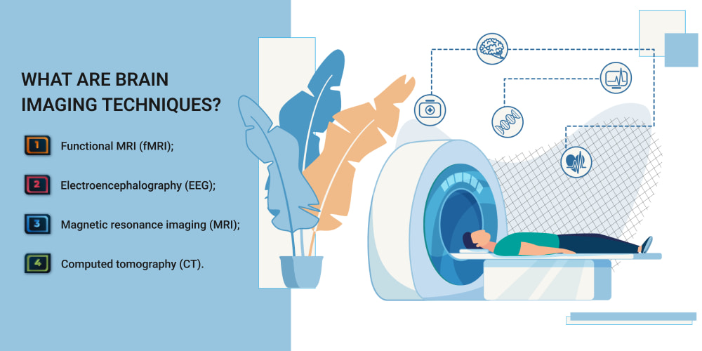
Brain imaging is essential for detecting any abnormalities and conditions in the human brain. The techniques help to provide an accurate picture of the structure of brain functions and activity. Healthcare professionals can monitor brain function and support various surgical planning. Here are the main techniques and their characteristics:
- Functional MRI (fMRI). Thanks to fMRI, it is possible to measure changes in blood flow and various active areas of the brain. It is ideal for studying brain function and cognitive activity. FMRI is often used in neurosurgery to obtain high resolution when examining the brain.
- Electroencephalography (EEG). Electrodes on the human scalp are used to record the electrical activity of the brain. It is used to diagnose epilepsy and various brain waves during the examination. This method is speedy, non-invasive, and ideal for analyzing brain activity. It is usually used in real-time.
- Magnetic resonance imaging (MRI). MRI relies on the use of strong magnetic fields to create detailed images of brain tissue. It is used to detect brain tumors and serious illnesses such as stroke. It has a high resolution and does not affect a person’s radiation.
- Computed tomography (CT). Such neuroimaging techniques as CT use X-rays. Usually, CT scans are used to diagnose traumatic brain injuries, skull fractures, and brain swelling. A significant advantage is the fast scanning time for quick examination and treatment.
Brain imaging with these tools allows for early detection of various diseases. Real-time monitoring of brain activity and its study is performed. It becomes possible to study cognitive habits during multiple seizures. Thanks to these tools, it is possible to quickly support the treatment management and surgical planning of brain tumors.
Functional MRI: A Breakthrough in Neurological Diagnosis
Neurological health plays a key role in a person’s quality of life. LoneStarNeurology provides support to patients and treats them with dedication by utilizing advanced technology and innovative treatment approaches. Thanks to functional MRI, scientists and doctors can visualize brain activity. Everything happens in real-time, and different structural images of the brain appear. It becomes possible to understand how different parts of the brain function.
The fMRI detects changes in oxygen levels in the brain when a specific part of the brain becomes active. When it needs more oxygen, this leads to increased blood flow, which fMRI can detect. Researchers and doctors can get all the information about some regions of the brain. Functional MRI has changed the diagnosis of neurological disorders. Here are its key capabilities:
- It helps to determine which areas of the brain are affected and to restore lost function. This process is usually very relevant in the rehabilitation plan after a stroke.
- Detects seizures and identifies areas for surgical intervention in patients. Detecting epileptic seizures at early stages will help improve health.
- In Alzheimer’s disease, it monitors early functional changes. It is one of the best neuroimaging techniques for assessing memory-related brain areas.
- Excellent for assessing dysfunction in Parkinson’s disease and various mental disorders. It helps to detect altered brain activity in patients with disorders such as anxiety and stress.
FMRI helps to assess brain function and detect early brain diseases. It remains one of the most valuable diagnostic tools in neuroscience. Doctors can prescribe treatment based on this data and get a good result.
Electroencephalography (EEG): Real-Time Brain Activity Monitoring
Electroencephalography (EEG) is a non-invasive way to measure electrical activity in the brain. It can record brain waves in real-time. This process is ideal for diagnosing and monitoring various neurological conditions. Here are the main advantages of EEG in brain monitoring and research:
- Real-time data collection. Data collection provides instant feedback, which is a priority. Doctors can study and investigate the dynamics of brain function.
- High temporal resolution. Electroencephalography detects the most minor changes in a very short time for fast analysis.
- Non-invasive and safe. It is ideal for different age groups due to the elimination of radiation and strong magnetic fields.
- Portability and cost-effectiveness. It is an affordable method for research and is used in various clinical and research settings.
EEG has a significant impact on the study of neurological disorders. In the diagnosis and monitoring of some brain-related diseases, EEG is the best solution. Here are the prominent examples of where it is best and how it works:
- Epilepsy. Electroencephalography detects seizure activity through electrical patterns. It can identify seizure types and plan treatment strategies.
- Sleep disorders. It is used to analyze changes in brain waves during different stages of sleep. It can detect various syndromes, especially restless legs syndrome.
- Assessment of traumatic brain injury. It helps to assess brain function in unconscious patients. Ideal for patients with coma and traumatic brain injury.
- Alzheimer’s disease. Can detect altered brain waves and evaluate effective treatment models.
- Migraine. Electroencephalography is the best for studying abnormal activity in the brain. It can give doctors an understanding of the mechanisms of pain perception in the brain.
How Brain Imaging Techniques Aid in Diagnosing Neurological Disorders
Neuroimaging techniques are essential tools for diagnosing a patient’s neurological condition. They are revolutionizing neurology and providing accurate, data-driven information. Doctors, in turn, have time to provide individualized treatment at early stages. This is how imaging contributes to a number of neurological diseases:
- Diagnosis of Alzheimer’s disease. Usually, functional MRI is used to scan specific parts of the brain to detect the disease. MRI and CT are essential for tracking brain atrophy. They also investigate its reduction in certain areas. MRIs allow for better treatment planning based on the findings.
- Detection of Parkinson’s disease. MRI is usually used to visualize the neurons that produce Dopamine. MRI can detect structural changes in the brain that distinguish this disease from other disorders. Brain imaging supports a more accurate and earlier diagnosis of treatment.
- Detection of multiple sclerosis. MRI reveals lesions of the brain and spinal cord. MRI helps to identify active inflammation and track the progression of the disease before treatment.
Continuous development of advanced imaging technologies contributes to improved research. It becomes possible to make an automated neurological diagnosis. Due to the detection of various abnormalities in the early stages, an individualized treatment plan is drawn up. Imaging and detection of brain conditions is possible due to the suitable resolution.
The Role of Brain Imaging in Personalized Treatment Plans
Brain imaging has a significant impact on individualized treatments. It plays a crucial role in building these data-driven strategies. Visualization helps doctors understand the entire basis of the structure and function of the patient’s brain. Technology allows doctors to tailor treatment to the patient’s condition. Here’s how imaging helps in choosing the proper treatment:
- With MRI or CT scans, stroke patients receive the best drug based on data. MRI helps to identify different conditions and processes in the brain. In multiple sclerosis, MRI monitors the progression of the disease.
- Functional MRI is integral for information about brain activity. Doctors can choose the proper medication for mental disorders such as depression or schizophrenia. Special instruments will be used to measure dopamine and serotonin for a suitable treatment plan.
- FMRI and EEG help to customize the proper treatment for epilepsy. They examine the functional areas of the brain and maintain deep brain stimulation. Based on the data, the patient can receive an individualized treatment plan.
Thanks to brain imaging, doctors can use this data to guide and brain scan analyses. Artificial intelligence helps to detect data invisible to human eyes. Monitoring the effects of various drugs in real time helps to adjust treatment. Integration of data with imaging and genetics leads to individualized drug prescriptions.
The Future of Brain Imaging in Neurological Diagnosis
The future of imaging in neurological diagnosis promises to be even better and more accurate. Continuous improvement allows for high-speed and non-invasive diagnostics. AI makes it possible to detect various abnormalities automatically. Doctors get quick image analysis to shorten the diagnosis. Equally important is prognostic analytics, which predicts the progression of multiple diseases. Brain imaging provides clear, detailed images with early detection of various changes in the brain. Studies show a better distinction between neurological disorders.
Future innovations include high-quality image processing devices. They will provide continuous monitoring of brain activity in real-time. It will be possible to conduct home diagnostics for conditions such as poor sleep, epilepsy, and migraines. Researchers are developing special scanners that will improve neurological diagnosis. The future will enable faster, more personalized treatment for any patient. Medicine will become even more accessible thanks to the use of artificial intelligence.
 (No Ratings Yet)


I've given up... the stress her office staff has put me through is just not worth it. You can do so much better, please clean house, either change out your office staff, or find a way for them to be more efficient please. You have to do something. This is not how you want to run your practice. It leaves a very bad impression on your business.
Please, leave your review
Write a comment: