

When neurologic symptoms appear, many patients aren’t sure what to expect next. Your primary care doctor might mention imaging, a friend might talk about getting an EEG test, and online searches often create more confusion than clarity. Patients frequently ask which brain scan they need and why one test is chosen over another. The choice between brain MRI vs CT or deciding whether an EEG is necessary can feel overwhelming without clear guidance.
Neurologists follow a structured decision-making process when ordering tests – they don’t just pick one at random. Each option – MRI, CT, or EEG – serves a specific purpose and answers different questions about what’s happening in your brain. Safety, urgency, your symptom pattern, and the type of neurologic imaging needed all play a role in the decision.
This article explains MRI, CT, and EEG in clear terms. It outlines how neurologists think, what each test does best, and how your symptoms guide testing choices. By the end, you’ll better understand why a specific test may be recommended and how to move through the process with confidence.
How Neurologists Decide Between Brain MRI and CT Scan
The choice between brain MRI vs CT often comes down to timing, detail, and how urgent your situation is. Both scans look at brain structure, but they work in different ways and at different speeds.
Key differences at a glance:
| Feature | CT Scan | MRI |
| Technology | X-rays (Radiation) | Magnetic Fields & Radio Waves |
| Duration | 2–5 minutes | 30–60 minutes |
| Best For | Acute bleeding, bone fractures | Soft tissue, tumors, nerve damage |
| Comfort | Open, fast, less noisy | Enclosed space, very loud |
A CT scan uses X-rays to create cross-sectional images of your brain. It’s fast, widely available, and excellent at detecting bleeding, fractures, and major structural changes. In emergency settings, speed is critical. When a patient arrives with sudden confusion, head trauma, or a possible stroke, a CT scan is often the first step because it can be completed within minutes.
An MRI uses magnetic fields and radio waves instead of radiation. It takes longer to perform, but provides much more detailed images of brain tissue, nerves, and subtle abnormalities. MRI is better for conditions that develop over time, such as chronic headaches, seizures, or suspected inflammatory disease. When deciding which brain scan is right for you, doctors balance the need for speed against the need for high-resolution detail.
Neurologists match the scan to the question they need answered. If the concern is active bleeding or a skull injury, CT usually comes first. If the concern involves nerve pathways, small lesions, or complex tissue changes, MRI offers greater clarity.
When MRI Is the Best Choice for Brain Imaging
MRI plays a crucial role in neurologic imaging when detail matters more than speed. It’s exceptional at showing differences in soft tissues, making it the preferred test for many neurologic conditions.
Patients with suspected multiple sclerosis often get an MRI because it can detect small plaques in the brain and spinal cord that CT scans might miss. Brain tumors, especially in early stages, also appear better on MRI because it highlights the contrast between normal and abnormal tissue.
Unexplained headaches that won’t go away despite treatment may lead your neurologist to order an MRI to rule out structural causes. MRI can also detect prior small strokes, inflammation, or subtle changes associated with memory loss. In these situations, neurologists prefer MRI because it provides the kind of answers that guide long-term care decisions.
When CT Scan Is the Most Practical Option
CT scans remain essential in neurology, particularly when time and access are critical. In many emergencies, CT is the most practical choice.
When a stroke is suspected, a CT scan helps determine whether there’s bleeding in the brain. This information shapes immediate treatment decisions and can be life-saving. Head trauma evaluation relies heavily on CT to detect fractures, bleeding, or swelling that may require urgent intervention. Patients with sudden, severe headaches often get a CT scan to rule out a brain hemorrhage.
CT scans are also more accessible in many settings and cost less than an MRI. For patients who can’t tolerate enclosed spaces or have implanted devices that aren’t MRI-compatible, CT offers a reliable alternative.
When Is an EEG Test Recommended Instead of MRI or CT?
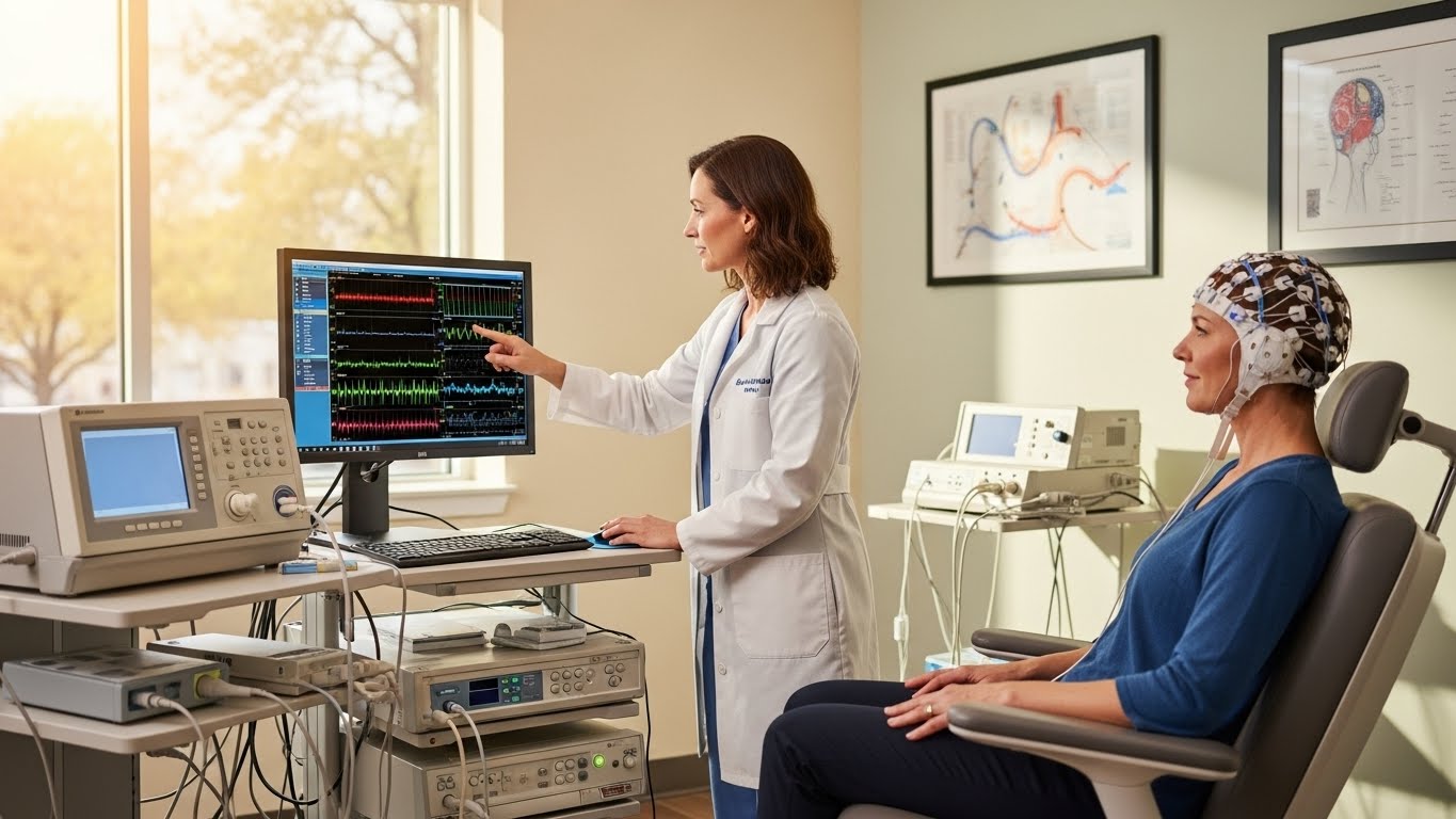
Unlike MRI and CT, an EEG test doesn’t look at brain structure. Instead, it records your brain’s electrical activity. This fundamental difference determines when and why neurologists recommend it.
EEG is commonly used to evaluate seizures. When a patient has episodes of losing awareness, shaking, or unexplained spells, an EEG test helps detect abnormal electrical patterns that suggest epilepsy or seizure activity. MRI or CT may appear completely normal in these cases because structural changes aren’t always present – the problem lies in how the brain is functioning, not how it looks.
An EEG test also helps evaluate fainting episodes, sleep disorders, and certain types of confusion. In hospital settings, EEG monitoring helps assess altered mental status or suspected nonconvulsive seizures.
Neurologists often use EEG alongside imaging tests. MRI or CT may rule out tumors or bleeding, while an EEG clarifies how your brain is functioning electrically. When you understand MRI, CT, and EEG explained together, it becomes clear that these tests complement rather than replace each other.
Situations where an EEG test provides added value include:
- Episodes of confusion without clear triggers
- Suspected focal or generalized seizures
- Blackouts with rapid recovery
- Abnormal movements during sleep
- Cognitive changes with fluctuating alertness
- Monitoring how well seizure treatment is working
Neurologists may recommend an EEG test when your clinical history suggests abnormal brain signaling, even if MRI or CT results look normal.
How Symptoms Determine the Right Brain Scan
Your symptoms guide every testing decision in neurology. Rather than ordering the same test for every patient, neurologists examine patterns and timing to determine which brain scan provides the most useful information.
Headaches that start suddenly and feel severe raise concern for bleeding and often lead to a CT scan first. Chronic headaches without red flags may prompt an MRI to evaluate deeper brain structures. Memory changes typically develop gradually and often call for an MRI to evaluate brain tissue and look for signs of prior injury.
After a head injury, a CT scan evaluates acute damage, while an MRI may follow if symptoms persist. Each symptom pattern points neurologists toward the most informative tool.
Symptom patterns that strongly influence scan choice include:
- Sudden neurologic problems within hours (weakness, speech changes, confusion)
- Gradual memory or personality changes over weeks or months
- Recurrent brief episodes of altered awareness
- Persistent balance problems or unexplained dizziness
- New neurologic symptoms after head trauma
- Vision loss affecting one or both eyes
Neurologists also evaluate how your symptoms change over time. Sudden onset symptoms raise different concerns than slow, progressive changes. The presence of fever, infection, recent injury, or medication changes also influences test selection. By combining symptom timing, severity, and related signs, neurologists reduce unnecessary testing and focus on studies most likely to produce useful answers.
The brain MRI vs CT question is one of the most common in neurology. Both are valuable tools, but they’re designed for different situations.
Think of it this way: CT is like taking a quick snapshot – it’s fast, convenient, and great for capturing the big picture. MRI is like using a professional camera with special lenses – it takes more time, but the detail and clarity are far superior.
For emergencies like suspected stroke, head trauma, or sudden severe headaches, CT wins because time matters. For conditions that develop over time – like multiple sclerosis, brain tumors, or chronic headaches – MRI provides the detail needed to make an accurate diagnosis.
Many patients end up getting both types of scans at different points in their care. You might get a CT in the emergency room to rule out immediate danger, then follow up with an MRI later for a more detailed evaluation.
Choosing a Trusted Neurology Clinic for Brain Imaging
Selecting the right clinic matters as much as selecting the right test. You benefit from care teams that combine experience, advanced technology, and clear communication.
Board-certified neurologists bring specialized training in interpreting imaging results within the context of your specific symptoms. Advanced scanners improve image quality and can shorten exam times. Access to EEG test specialists ensures accurate testing and interpretation.
A strong neurology clinic creates a personalized imaging plan based on your symptoms, medical history, and concerns. Clear explanations reduce anxiety and help you feel involved in your care decisions.
If you’re experiencing neurologic symptoms and need answers, scheduling a consultation lets you receive a tailored recommendation on which brain scan is right for you and move forward with confidence, supported by expert care.
 (No Ratings Yet)


I've given up... the stress her office staff has put me through is just not worth it. You can do so much better, please clean house, either change out your office staff, or find a way for them to be more efficient please. You have to do something. This is not how you want to run your practice. It leaves a very bad impression on your business.
Please, leave your review
Write a comment: